Rare IgG4-related GI disease often mistaken for stomach tumors
Study: Defining features of disorder could help avoid unnecessary surgeries
Written by |
Researchers in Japan have described the clinical features of a large group of people with IgG4-related disease (IgG4-RD) affecting the gastrointestinal tract, shedding new light on this poorly characterized manifestation of the condition.
In addition to four previously reported hallmark tissue features, IgG4-related gastrointestinal disease (IgG4-GID) appeared almost exclusively in the upper gastrointestinal tract and most often presented as ulcers or tumor-like masses.
Because these mass-like lesions closely resembled stomach tumors, many patients underwent surgery before the underlying IgG4-RD was recognized. Most lesions, however, were reduced with glucocorticoids — the first-line IgG4-RD treatment — and/or stomach acid-reducing medications.
“The current study not only reinforces previous findings but also provides new insights into the clinical and [tissue-related] spectrum of IgG4-GID,” researchers wrote.
While additional cases are needed to draw a clearer picture, the team emphasized that defining the characteristic features of IgG4-GID can help clinicians exclude mimicking conditions and ultimately “avoid unnecessary surgeries.”
The study, “Clinical manifestations of immunoglobulin G4‑related gastrointestinal disease: a nationwide multicenter retrospective study,” was published in the Journal of Gastroenterology.

GI manifestations of IgG4-RD reported in small number of cases
In IgG4-RD, immune cells — particularly those that produce IgG4-type antibodies — infiltrate tissues, causing inflammation and scarring that leads to swelling and tumor-like masses in one or more organs. Over time, this immune-driven damage can disrupt normal organ function and lead to a range of IgG4-RD symptoms.
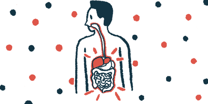
While the pancreas, salivary glands, lungs, and kidneys are well-established sites of involvement, gastrointestinal, or GI, manifestations have been reported in a small number of cases.
While IgG4-GID is “gradually being recognized as a subgroup of IgG4-RD, the disease concept has not yet been established,” the researchers wrote.
To address this gap, a team of researchers in Japan collected and analyzed data from 37 suspected IgG4-GID cases identified across 35 institutions in the country. Eligible cases dated back as far as 2001 and included those with gastrointestinal lesions showing infiltration of IgG4-positive immune cells, other tissue features suggestive of IgG4-RD, or lesions that improved with standard glucocorticoid treatment.
In this study, no definitive cases of IgG4-GID were identified in the [whole] small intestine or colon [large intestine]; however, further studies are needed to determine whether lesions occur in these areas.
A total of 18 patients were classified as “definite” or “highly likely” to have IgG4-GID based on the presence of any of four tissue features previously reported to be associated with IgG4-GID, with or without many IgG4-positive immune cells.
These patients, with a median age of 69.5 years and a predominance of females (66.7%), were included in the final analysis.
All cases involved the upper gastrointestinal tract: 11 affected the stomach, five involved the duodenum (the first section of the small intestine), one affected the esophagus (the food pipe), and one involved both the stomach and duodenum.
“In this study, no definitive cases of IgG4-GID were identified in the [whole] small intestine or colon [large intestine]; however, further studies are needed to determine whether lesions occur in these areas,” the researchers wrote.

Ulcers most common abnormality observed during endoscopy
Ulcers were the most common abnormality observed during endoscopy, affecting 38.9% of participants. Most of these patients (57.1%) had multiple ulcers, while 42.9% had a single ulcer. Endoscopy is a medical procedure in which a long, thin tube equipped with a light and a camera is inserted through the mouth or anus for diagnostic or therapeutic purposes.
One-third of participants showed lump-like lesions beneath the stomach lining. Because these resembled stomach tumors, all of these patients underwent surgery before IgG4-RD was recognized as the actual cause.
Two other patients underwent surgery, one who had a small erosion in the esophagus, and another who showed no abnormality on endoscopy but was found to have stomach wall thickening during surgery.
Most of the 18 patients (77.8%) also had IgG4-RD involving other organs, most commonly the pancreas (44.4%), followed by the bile ducts (22.2%) and the salivary or tear glands (16.7%). The bile ducts are the tubes that transport the digestive fluid bile from the liver to the small intestine.
When examining tissue samples, the researchers identified the four previously reported inflammatory patterns that appear to characterize IgG4-GID. These features included inflammation extending deep into the GI tract’s muscle layer in a striped or layered pattern and antibody-producing immune cells packed densely in the deeper parts of the stomach or duodenal lining.
The other two features were tumor-like inflammatory growths beneath the inner lining of the GI tract and inflammation of the Brunner’s glands, which are located in the duodenum.
Treatment varied depending on the type of GI involvement. Overall, nearly half of the confirmed cases (44.4%) underwent surgery, primarily because their lesions closely resembled tumors. Others were treated medically.
One person (5.6%) received glucocorticoids alone, three (16.7%) were given acid-reducing medications for ulcers, and four (22.2%) received a combination of the two. All but one responded to therapy, with no recurrences observed during follow-up. The single non-responder had an old ulcer scar rather than active disease.
“In this nationwide multicenter study, we identified a group of cases likely to be definitive of IgG4-GID,” and highlighted its “characteristic [tissue] findings and various endoscopic features,” the researchers wrote. “However, many aspects of their clinical features remain unclear, and further accumulation of cases is required for future studies.”