
New tool assesses IgG4-RD organ damage, disease activity
Researchers develop disease activity scoring system based on scan results
Written by |
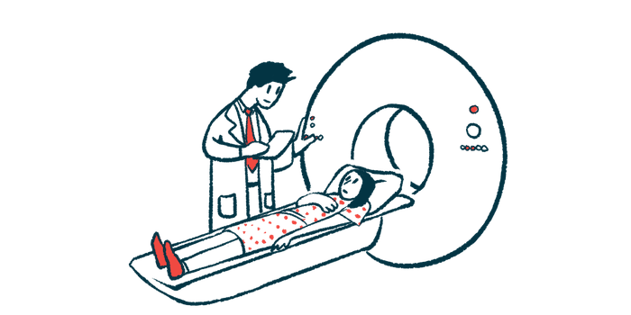
A new imaging technique shows promise for assessing disease activity and organ involvement in immunoglobulin G4-related disease (IgG4-RD).
Compared with biopsy results, the [18F]FAPI PET/CT scan was 100% accurate in detecting lesions in various organs, including the lung, liver, muscles, pancreas, and kidney.
The researchers developed a disease activity scoring system based on scan results that correctly distinguished active from inactive disease with 93% accuracy. They said the system has “the potential to enhance disease stratification in these patients, thereby improving diagnostic accuracy and informing therapeutic decision-making in clinical practice.”
The study, “[18F]FAPI PET/CT-based scoring systems for patient assessment in IgG4-related disease,” was published in RMD Open.
IgG4-RD is a disorder in which the immune system drives inflammation and fibrosis (scarring) in various organs. It’s characterized by elevated blood levels of the IgG4 antibody subtype and the build-up of IgG4-producing immune plasma cells in affected tissues.

Limitations of current imaging methods
One imaging approach used in IgG4-RD is [18F]F-fluorodeoxyglucose PET/CT, a hybrid technique that combines PET and CT scans. It uses 18F-fluorodeoxyglucose, a radioactive tracer of glucose (cells’ main energy source), to measure a lesion’s metabolic activity and assess its size and shape. Active lesions typically show increased tracer uptake, and the degree of uptake reflects the level of inflammation.
Still, it has limitations. Similar patterns of increased uptake are observed in cancers, infections, and other autoimmune conditions. And in IgG4-RD lesions built on fibrosis, metabolic activity may be reduced, leading to false-negative results.
A newer tracer, 18F-fibroblast activation protein inhibitor (FAPI), binds to FAP, an enzyme involved in activating fibroblasts, the cells responsible for scarring in IgG4-RD. [18F]FAPI PET has shown greater sensitivity than conventional imaging for detecting active fibrosis, “particularly in organs like the pancreas and salivary glands, which are commonly affected in IgG4-RD,” the researchers wrote.
Prior research demonstrated that [18F]FAPI PET/CT can detect organ involvement in IgG4-RD even in the absence of symptoms. And the total lesion FAPI uptake, a measure of tracer uptake across all lesions, was significantly associated with IgG4-RD activity.
To date, however, no study has created a standardized method for using this technique to measure disease activity in IgG4-RD. With this in mind, a team of researchers in China retrospectively analyzed demographic, clinical, and imaging data from 85 adults with IgG4-RD (67.1% men; average age 57) and 10 healthy individuals (50% men; average age 62.4).
To determine whether an organ exhibited abnormal tracer uptake, the researchers established thresholds based on data from healthy individuals. Any uptake above this threshold was considered abnormal for that organ.
Biopsy, in which a tiny tissue sample is collected for analysis under a microscope, confirmed 64 IgG4-RD-associated lesions across patients. Of these, 58 were correctly identified as positive by the [18F]FAPI PET/CT scan thresholds, while six were classified as negative, yielding an overall diagnostic accuracy of 85.9%.
The scan’s accuracy was 100% for lesions in certain regions of the body, including the pancreas, salivary glands, lungs, skeletal muscle and skin, liver, kidney, and prostate. By comparison, accuracy was lowest (below 70%) in the neck, armpit, and chest lymph nodes (immune structures).
Of the 85 patients, 59 had active IgG4-RD and 26 had inactive disease, as assessed by the standard IgG4-RD Responder Index. Blood IgG4 levels were significantly higher in the active group (7.6 g/L vs. 2.4 g/L).
The active group showed significantly higher values in three image-derived measurements. Across numerous organs, tracer uptake was higher in active disease, including in the thyroid gland, salivary glands, lymph nodes, lungs, arteries, pancreas, kidney, prostate, and heart.
The team noted that higher uptake in the heart, a previously overlooked area, was observed in active disease. This suggests the potential accumulation of IgG4-positive plasma cells in the heart muscle, warranting consideration in clinical management.
Researchers then developed an [18F]FAPI PET/CT activity score (FPAS-IgG4) based on FAPI uptake intensity and structural abnormalities at each organ or site. Overall, IgG4-RD patients with active disease had a significantly higher FPAS-IgG4 score than those with inactive disease (5.6 vs. 1.7 ).
A statistical analysis found that FPAS-IgG4 was strongly associated with active IgG4-RD, with a sensitivity, or the ability to correctly identify patients with active disease, of 93.2%. Its specificity, or the ability to correctly rule out patients without active disease, was 73.1%.
Among the laboratory markers examined, higher blood IgG4 levels were significantly associated with higher FPAS-IgG4 scores.
“The findings from our study indicate that [18F]FAPI PET/CT may serve as an effective tool for monitoring disease activity in patients with IgG4-RD,” the team concluded. “The newly developed FPAS-IgG4 activity scoring system may be beneficial in stratifying disease in these patients.”


Leave a comment
Fill in the required fields to post. Your email address will not be published.